
C57BL/6N-Pdcd1tm3(PDCD1)Bcgen Tgfbr2tm3(TGFBR2)Bcgen/Bcgen • 111890
| Product name | B-hPD-1 plus/hTGFBR2 mice |
|---|---|
| Catalog number | 111890 |
| Strain name | C57BL/6N-Pdcd1tm3(PDCD1)Bcgen Tgfbr2tm3(TGFBR2)Bcgen/Bcgen |
| Strain background | C57BL/6N |
| NCBI gene ID | (Human) |
| Aliases | PD1; PD-1; CD279; SLEB2; hPD-1; hPD-l; hSLE1; ADMIO4; AIMTBS; AAT3; FAA3; LDS2; MFS2; RIIC; LDS1B; LDS2B; TAAD2; TBRII; TBR-ii; TGFR-2; tbetaR-II; TGFbeta-RII |
Gene targeting strategy for B-hPD-1 plus/hTGFBR2 mice.
Human PD-1 gene encoding the extracellular region and mouse PD-1 gene encoding the transmembrane and cytoplasmic region were inserted after the initiation codon ATG of mouse PD-1 gene in B-hPD-1 plus/hTGFBR2 mice.
A chimeric CDS that encodes mouse TGFBR2 signal peptide, human extracellular domain, mouse Tgfbr2 transmembrane and cytoplasmic domain, followed by mouse 3’UTR-STOP is inserted right after mouse the exon 2 of Tgfbr2 to replace the exon 2 of mouse Tgfbr2 gene. The chimeric TGFBR2 protein expression will be driven by endogenous mouse Tgfbr2 promoter, while mouse Tgfbr2 gene transcription and translation will be disrupted.
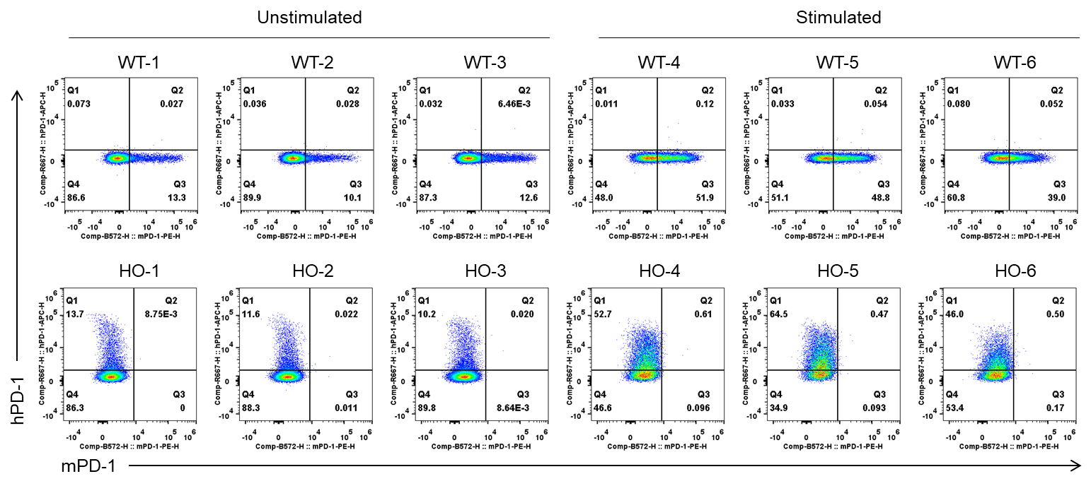
Strain specific PD-1 expression analysis in wild-type mice (WT) and homozygous (HO) B-hPD-1 plus/hTGFBR2 mice by flow cytometry. Splenocytes were collected from wild-type C57BL/6JNifdc mice (+/+) and homozygous B-hPD-1 plus/hTGFBR2 mice after stimulated with anti-mouse CD3ε antibody (7.5 μg, i.p.) in vivo for 24 hrs (female, 10-week-old, n=3) or not. Protein expression was analyzed with anti-mouse PD-1 antibody (Biolegend, 109104), and anti-human PD-1 antibody (Biolegend, 329904), by flow cytometry. Mouse PD-1 was detectable in wild-type C57BL/6JNifdc mice, but not in the homozygous B-hPD-1 plus/hTGFBR2 mice. Human PD-1 was exclusively detectable in B-hPD-1 plus/hTGFBR2 mice.
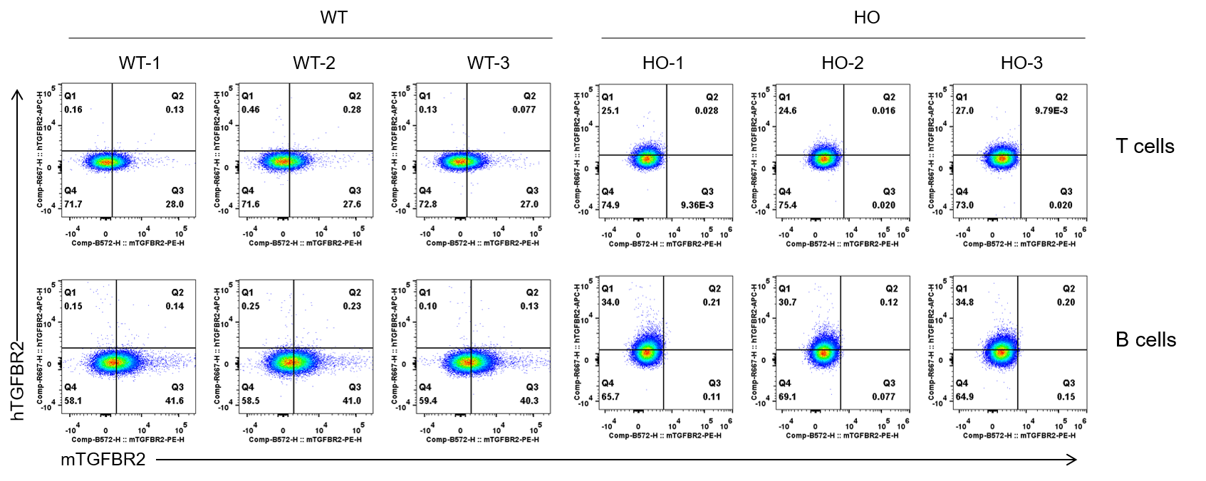
Strain specific TGFBR2 expression analysis in wild-type (WT) C57BL/6JNifdc mice and homozygous (HO) B-hPD-1 plus/hTGFBR2 mice by flow cytometry. Splenocytes were collected from wild-type C57BL/6JNifdc mice (+/+) and homozygous B-hPD-1 plus/hTGFBR2 mice (female, 10-week-old, n=3). Protein expression was analyzed with anti-mouse TGFBR2 antibody (R&D, FAB532P), and anti-human TGFBR2 antibody (Biolegend, 399705) by flow cytometry. Mouse TGFBR2 was detectable in wild-type C57BL/6JNifdc mice, but not in the homozygous B-hPD-1 plus/hTGFBR2 mice. Human TGFBR2 was exclusively detectable in B-hPD-1 plus/hTGFBR2 mice.
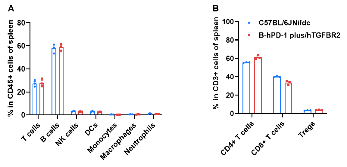
Frequency of leukocyte subpopulations in spleen by flow cytometry. Splenocytes were isolated from wild-type C57BL/6JNifdc mice and homozygous B-hPD-1 plus/hTGFBR2 mice (female, 10-week-old, n=3). A. Flow cytometry analysis of the splenocytes was performed to assess the frequency of leukocyte subpopulations. B. Frequency of T cell subpopulations. Frequencies of T cells, B cells, NK cells, dendritic cells, monocytes, macrophages, neutrophils, CD4+ T cells, CD8+ T cells, and Tregs in B-hPD-1 plus/hTGFBR2 mice were similar to those in C57BL/6JNifdc mice. The frequency of leukocyte subpopulations in blood and lymph nodes of B-hPD-1 plus/hTGFBR2 mice were also comparable to wild-type C57BL/6JNifdc mice (Data not shown). Values are expressed as mean ± SEM. Significance was determined by two-way ANOVA test. *P < 0.05, **P < 0.01, ***p < 0.001.